How We Spot Glaucoma Before You Notice Vision Loss: The Science of Digital Eye Exams
Glaucoma is a progressive ocular condition that can damage your sight so quietly that visual changes go unnoticed until permanent damage occurs. For individuals and families seeking vision care guidance, prescription eyewear, or contact lenses in West Lebanon, NH, and surrounding communities, understanding how this condition behaves is vital. Because early optic nerve damage does not produce pain or change central clarity, self-diagnosis is a high-risk strategy. Instead, scheduling a modern digital eye exam for glaucoma represents your most effective shield against silent vision loss. By utilizing advanced imaging and measurement technologies, eye care professionals can identify microscopic structural changes in the eye years before you notice any visual deficits, allowing for early intervention that preserves your daily independence.
What is Glaucoma and Why is It Called the Silent Thief?
Glaucoma is a group of progressive eye conditions characterized by damage to the optic nerve. This nerve acts as the primary transmission cable of the visual system, carrying millions of electrical impulses from the retina to the brain to be translated into images. When these delicate nerve fibers are damaged, communication between your eyes and brain is permanently disrupted.
In most cases, this damage is associated with elevated intraocular pressure. The eye constantly produces a clear fluid called aqueous humor to nourish its internal structures. This fluid must drain out at the same rate it is produced to maintain a healthy pressure. If drainage channels become clogged or sluggish, fluid builds up, causing internal pressure to rise and placing physical stress on the optic nerve fibers, leading to their decay.
Glaucoma is called the silent thief of sight due to the complete lack of glaucoma early symptoms in open-angle glaucoma, its most common form. It typically begins by damaging nerve fibers responsible for peripheral vision. Because central vision remains clear and the brain naturally compensates for small blind spots, most people are unaware of any shrinkage in their visual field until irreversible damage has already occurred.

The Science of a Digital Eye Exam for Glaucoma
Modern optometry has moved far beyond basic visual acuity charts to detect complex nerve conditions. A comprehensive digital eye exam for glaucoma combines several highly specialized measurement systems to analyze both the physical structure of your eye and its functional performance. This multi-layered approach provides an exceptionally accurate baseline map of your ocular health.
Checking Eye Pressure with Precision
Checking eye pressure, clinically known as tonometry, measures the resistance of your cornea to light physical contact. Rebound tonometry utilizes a microscopic, ultra-lightweight probe that contacts the cornea for a fraction of a second. This measurement is completely painless, does not require numbing drops, and is comfortable and straightforward for adults of all ages.
Because intraocular pressure fluctuates naturally, establishing a consistent record over time is critical. A single high pressure reading does not automatically indicate glaucoma, just as a normal reading does not guarantee you are free of the disease; it is one piece of a larger diagnostic puzzle.
Mapping the Optic Nerve with Optical Nerve Imaging
Optical nerve imaging allows clinicians to observe the physical structure of the optic nerve directly. The primary technology, Optical Coherence Tomography (an OCT scan), is a non-invasive imaging test that uses light waves to take high-resolution, cross-sectional pictures of your retina and optic nerve head.
Working similarly to ultrasound but using light waves, this test captures data in a matter of seconds while you look at a target light. The resulting digital images measure the thickness of your retinal nerve fiber layer with extreme accuracy. Because glaucoma thins this layer as it progresses, the scan detects microscopic changes years before any actual vision loss shows up on functional tests.
Evaluating Visual Function with a Visual Field Test
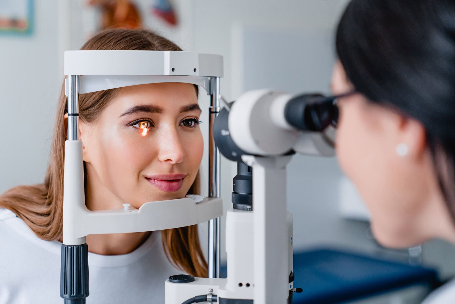
A visual field test (perimetry) evaluates functional performance by measuring your peripheral sensitivity to ensure no blind spots have developed.
During this computerized exam, you rest your chin in a bowl-shaped instrument and look at a central target light, pressing a button each time you detect a faint flash of light in your peripheral field. The computer maps your visual field, comparing results to age-matched healthy baselines. This verifies if any structural nerve thinning detected on an OCT scan is actively impacting your functional vision.
How Glaucoma is Diagnosed and Monitored
A clinical diagnosis is never made based on a single measurement or scan. Understanding how glaucoma is diagnosed requires looking at the synthesis of all structural and functional data collected during your evaluation. The independent doctor of optometry will evaluate your eye pressure, corneal thickness, the drainage angle of your eye, the physical appearance of your optic nerve head, and your visual field map.
Evaluating the eye's internal drainage angle determines if you are at risk for angle-closure glaucoma, a rapid form of the condition. Measuring corneal thickness via pachymetry is also essential, as thin corneas can cause standard tonometers to underestimate actual intraocular pressure.
Once diagnosed, the primary goal is to prevent glaucoma vision loss. Since glaucoma damage is irreversible, treatment focuses on halting progression by lowering intraocular pressure to a safe level, typically using daily prescription eye drops to reduce fluid production or improve drainage. Regular monitoring through repeated digital exams verifies that the treatment is successfully keeping your eye pressure stable.
Why Regular Vision Health Monitoring Matters After 40
The risk of developing glaucoma increases significantly as you age. While the condition can affect anyone, individuals over the age of forty should prioritize annual visual health evaluations. Several key factors can elevate your risk, including:
- Age: The biological efficiency of the eye's drainage systems naturally declines over time, making older adults more vulnerable to fluid buildup.
- Family History: Having a parent or sibling with glaucoma significantly increases your likelihood of developing the condition, suggesting a strong genetic link.
- Systemic Health Conditions: Chronic health issues like diabetes, high blood pressure, and cardiovascular disease can negatively impact blood flow to the optic nerve.
- High Prescriptions: Severe nearsightedness or farsightedness is structurally associated with anatomical variations that can predispose the eye to drainage issues.
Scheduling regular eye exams is your primary defense against silent progression. During these visits, updated prescriptions for eyeglasses can be determined to ensure your daily visual comfort, while the underlying health of your eyes is thoroughly assessed. Utilizing comprehensive eye care services guarantees that advanced diagnostic technologies are utilized to monitor your vision health continuously, allowing you to maintain your visual clarity and protect your quality of life as your eyes mature.

Frequently Asked Questions
Is a digital eye exam for glaucoma painful?
A digital eye exam for glaucoma is entirely painless and non-invasive, requiring no physical discomfort or down-time. Modern instruments like rebound tonometers and optical coherence tomography scanners utilize gentle light waves and micro-probes to measure eye structure and pressure in a matter of seconds without causing irritation.
Because these diagnostic tools do not touch the eye forcefully, most adults find the process highly comfortable. The entire evaluation is designed to be quiet, efficient, and stress-free, ensuring that patients can undergo regular screenings without any anxiety.
What are the primary glaucoma early symptoms?
In its most common form, open-angle glaucoma produces absolutely no early symptoms, pain, or warning signs. The gradual loss of peripheral vision occurs so slowly that the brain automatically compensates for the blind spots, leaving your central vision perfectly clear and giving no indication of a problem.
Because there are no obvious changes to alert you, the condition is virtually impossible to detect without specialized equipment. The only reliable way to identify early-stage glaucoma is through routine clinical evaluations that measure optic nerve structure and intraocular pressure.
How does checking eye pressure help detect glaucoma?
Checking eye pressure measures the fluid pressure inside the eye, which is a major, manageable risk factor for optic nerve damage. When the eye's natural drainage system becomes restricted, fluid builds up and increases pressure, placing physical stress on the delicate nerve fibers at the back of the eye.
Monitoring this pressure allows eye care professionals to identify elevated levels before they cause damage. However, since glaucoma can develop even with normal eye pressure, tonometry is always combined with detailed structural imaging of the optic nerve.
Why is an optical nerve imaging scan necessary?
An optical nerve imaging scan, such as an OCT, is necessary because it captures high-resolution, cross-sectional images of the retina to measure the thickness of the nerve fiber layer. Glaucoma causes these microscopic nerve fibers to thin years before any visible blind spots appear in your vision.
Measuring the physical thickness of this nerve fiber layer at a microscopic level allows clinicians to detect early signs of decay. This structural data serves as an invaluable baseline to monitor your eyes over time and detect changes.
How often should older adults get a visual field test?
Older adults with risk factors for glaucoma, such as a family history or elevated eye pressure, should undergo a visual field test at least once a year. For individuals already diagnosed with the condition, more frequent testing may be required to verify that their treatment is successfully halting the disease.
This test maps your peripheral sensitivity to ensure no blind spots have developed. Regular visual field assessments allow clinicians to track the functional health of your optic nerve and adjust your management plan to preserve your daily visual field.
Conclusion
Glaucoma remains a silent threat to vision health, but modern ophthalmic science has transformed how the condition is identified and managed. Relying on noticeable changes in your sight as a warning sign is a dangerous approach, as early optic nerve damage occurs entirely without symptoms. Prioritizing a regular digital eye exam for glaucoma is an effective, painless, and vital shield for your sight. By combining precise pressure measurements, advanced nerve fiber imaging, and functional peripheral mapping, eye care professionals can detect early indicators of pressure stress long before your vision is actively impacted. Taking a proactive approach ensures you preserve the visual clarity and independence necessary to enjoy life in the Upper Valley.
For individuals seeking additional information about this topic or professional vision care in West Lebanon, NH, Pro Optical provides eye exams, prescription eyewear, and contact lens services. Learn more at https://www.pro-optical.com/.